 Hablamos Español
Oligodendroglioma
Pure oligodendrogliomas are much less common than astrocytomas, making up only 5 – 20 percent of glial type brain tumors. Typically these come to attention in the fourth through sixth decades of life. They are treated in a similar manner with as complete a surgical resection as possible. Chemotherapy and radiation may also be used. Molecular genetics laboratory studies on the oligodendroglioma tissue demonstrate in some of these tumors a deletion of the short arm of the 1st chromosome and the long arm of the 19th chromosome. This is called a 1p 19q deletion and predicts better sensitivity to chemotherapy. However, just as with benign and malignant astrocytomas, it has unequivocally been established that patients treated with an aggressive primary surgical resection have longer survival rates.
In the example below, the patient presented to medical attention with no complaints other than new onset of seizures. Imaging studies demonstrated a low signal intensity abnormality with no appreciable enhancement with contrast. Surgery was performed, and the first portion of the resected tumor was sent for biopsy which demonstrated low-grade oligodendroglioma.
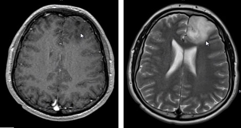
Two different sequences of MR imaging show the tumor which appears similar to brain in the T1 image on the left, with the abnormality better visualized on the T2 MR sequence on the right (arrows point to the posterior edge of the tumor).
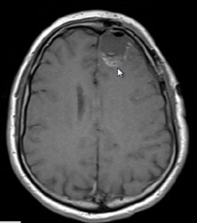
Owing to the difficulty in recognizing the brain tumor interface even by the pathologist studying biopsies from the margins of the tumor, it is my practice to get an early post-op MRI to look for possible residual tumor. This is evident in this post-op image (arrow points to tissue suspicious for residual tumor).
This patient therefore underwent an early second stage surgery for removal of this residual fragment.
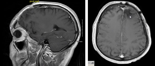
The above images are taken 7 years after the initial two-stage surgery, chemotherapy, and radiation. There is still no evidence of tumor recurrence. The patient continues to live an active life and work full time. He has had no more seizures.